No two weeks look the same at Neurospine International — and this one was a good reminder of just how varied our field really is. From facial pain to a silent vascular risk to a herniated disc that wouldn’t let go, here’s a look at three patients we helped this week.
When Facial Pain Has a Name: Trigeminal Rhizolysis
Few conditions cause pain as sharp and relentless as trigeminal neuralgia. Patients often describe it as an electric shock — triggered by something as simple as brushing teeth, chewing, or a gust of wind on the cheek. It stems from irritation or compression of the trigeminal nerve (cranial nerve V), most commonly at its root entry zone near the brainstem, though the pain itself is felt along one of its three branches — ophthalmic, maxillary, or mandibular — across the face.
For one patient this week, that pain had reached a breaking point. Rather than proceeding straight to open microvascular decompression, we opted for a percutaneous rhizolysis: accessing the trigeminal ganglion through the foramen ovale, a small opening at the base of the skull, under fluoroscopic guidance. A needle is advanced with millimetric precision — real-time imaging confirms the trajectory before any lesioning is done, since being even a few millimeters off target can mean the difference between relieving the right branch and missing it entirely.
Once positioned, the nerve fibers responsible for transmitting pain are selectively interrupted, while sparing as much normal sensory and motor function as possible. It’s a technique that rewards precision and patience — no open surgery, no craniotomy, and for many patients, relief is noticeable almost immediately. Recovery is typically measured in days, not weeks.
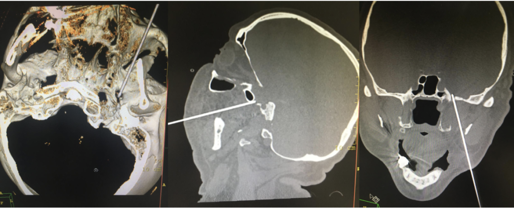
Clinical pearl: trigeminal neuralgia is a clinical diagnosis first — imaging helps rule out secondary causes like vascular compression or a mass lesion, but the classic paroxysmal, trigger-based pain pattern is what points us toward this diagnosis before the scan ever does.
The Aneurysm No One Knew Was There
Some of the most important findings in neurosurgery are the ones nobody was looking for. This week, a routine scan — ordered for an entirely different reason — uncovered an aneurysm at the anterior communicating artery (AComA), the vessel that connects the two anterior cerebral arteries at the front of the Circle of Willis.
It’s a notorious location for a reason. The AComA sits at a complex, high-flow junction, and aneurysms here account for a significant proportion of ruptured intracranial aneurysms — often presenting as subarachnoid hemorrhage when they do rupture, which carries serious morbidity and mortality. Finding one before it ruptures changes everything.
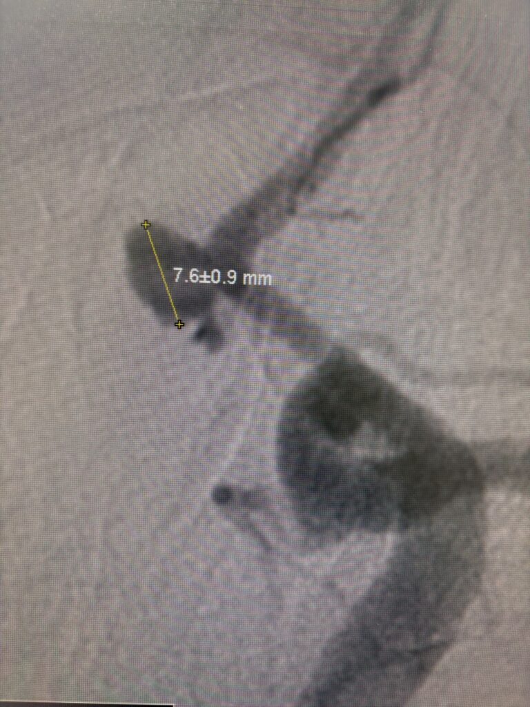
Our team performed a detailed diagnostic angiogram to map the aneurysm’s exact morphology — its neck width, dome size, and relationship to adjacent branches — all of which determine whether endovascular coiling, flow diversion, or surgical clipping is the safest route. Small anatomical details here carry real weight: a wide-necked aneurysm behaves very differently to treatment than a narrow-necked one, and the AComA’s proximity to critical perforating vessels means every millimeter of planning matters.
Clinical pearl: the AComA is one of the most common sites for intracranial aneurysms, and incidental discovery — as in this case — is becoming more frequent simply because we image the brain more often than we used to. It’s a strong argument for taking every incidental finding seriously, not dismissing it as background noise.
A Disc, a Nerve, and a Faster Way Back
Radicular leg pain can be one of the most frustrating things a patient lives with — sharp, shooting, often worse than the back pain that supposedly caused it. That’s because a herniated lumbar disc doesn’t just take up space; it compresses and inflames the nerve root exiting at that level, and the pain radiates along the path of that nerve, often all the way down to the foot.
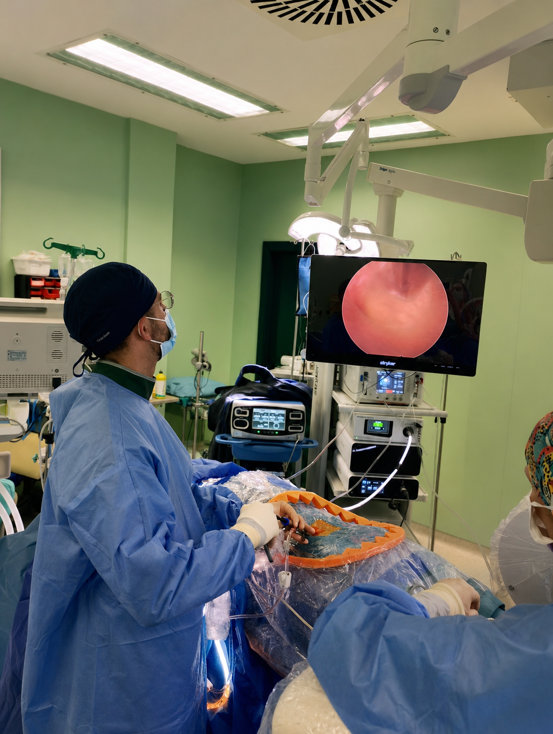
This week we performed an endoscopic lumbar microdiscectomy. Working through a small incision — often under a centimeter — a working channel is advanced under fluoroscopic guidance to the affected disc level. Through it, a high-definition endoscope gives a magnified, direct view of the herniated fragment and the nerve root it’s compressing, allowing us to remove the offending material with fine instruments while preserving the surrounding healthy disc, ligament, and bone.
Compared to traditional open discectomy, the endoscopic approach means less muscle disruption, less blood loss, and — for most patients — a dramatically shorter recovery. Many are up and walking the same day, with radicular pain often improving before they even leave the recovery room.
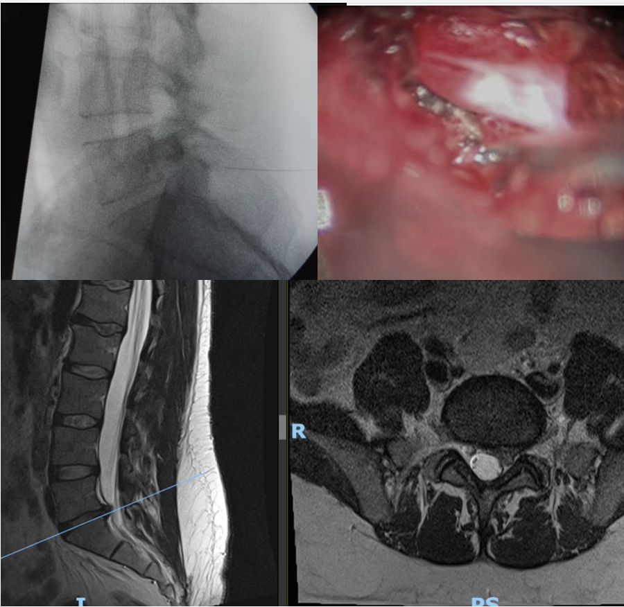
Clinical pearl: not every disc herniation needs surgery — most improve with conservative management. Surgery becomes the right call when pain is severe, unrelenting, or accompanied by neurological deficit, which was exactly the picture in this case.