
For many conditions of the brain and spine, the most direct route to the problem is not through the skull or the back. It is through the blood vessels.
Endovascular neurosurgery uses thin catheters passed through an artery, usually at the wrist or groin, and guided under real-time X-ray imaging to the vessels of the brain and spinal cord. From inside the vessel, specialists can see the circulation in fine detail and, in many cases, treat the disease without an open operation.
This article explains what endovascular techniques are, which conditions they are used for, and how they fit alongside conventional neurosurgery.
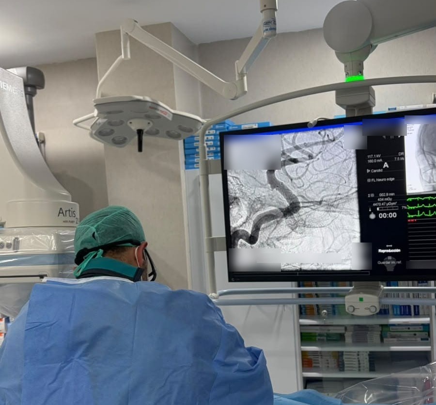
How an endovascular procedure works
The procedure takes place in an angiography suite, a room equipped with high-resolution X-ray imaging. After local anaesthesia (or general anaesthesia for most treatments), a small puncture is made in the radial artery at the wrist or the femoral artery at the groin.
A catheter is then guided through the arterial system to the vessel of interest. Contrast medium is injected and images are taken from several angles, often with 3D reconstruction. The result is a detailed map of the blood flow through the brain or spinal cord.
When treatment is needed, smaller microcatheters are guided further, into the diseased vessel itself, to deliver coils, stents, embolic agents or other devices.
Diagnosis: cerebral and spinal angiography
Digital subtraction angiography (DSA) remains the reference standard for studying the blood vessels of the central nervous system. CT and MR angiography are excellent first-line tests, but DSA offers higher resolution and shows blood flow over time, which matters for:
- Cerebral aneurysms: defining their exact shape, neck and relationship to nearby branches before choosing treatment.
- Arteriovenous malformations (AVMs): identifying feeding arteries, the nidus and draining veins.
- Dural arteriovenous fistulas: abnormal connections that can be hard to see on other scans.
- Spinal vascular malformations: a common hidden cause of progressive leg weakness and sphincter problems, often only confirmed by spinal angiography.
- Unexplained brain haemorrhage: to look for an underlying vascular cause.
- Tumour blood supply: to plan surgery on highly vascular lesions.
Treatment: what can be done from inside the vessel
Cerebral aneurysms
Many aneurysms, ruptured or unruptured, can be treated endovascularly. Options include:
- Coiling: soft platinum coils fill the aneurysm and exclude it from the circulation.
- Balloon- or stent-assisted coiling: for aneurysms with a wide neck.
- Flow diverters: fine-mesh stents placed across the aneurysm neck that redirect flow and allow the vessel wall to heal over time.
- Intrasaccular devices: implants placed inside the aneurysm sac for selected shapes.
A recent case from our team
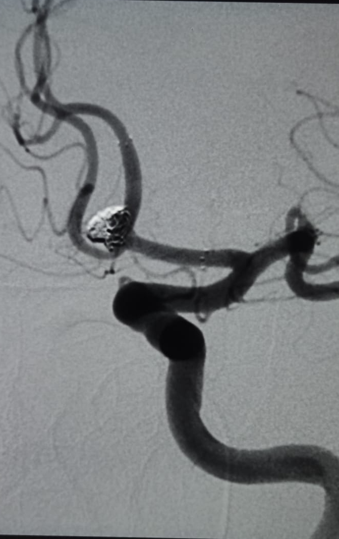
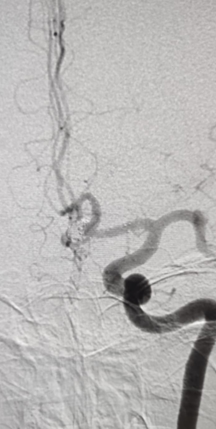
These control angiograms show the coil mass packed inside the aneurysm (bright white in the first image), sealing it off from the circulation while the surrounding cerebral arteries stay open. The aneurysm was treated through a small arterial puncture, without opening the skull.
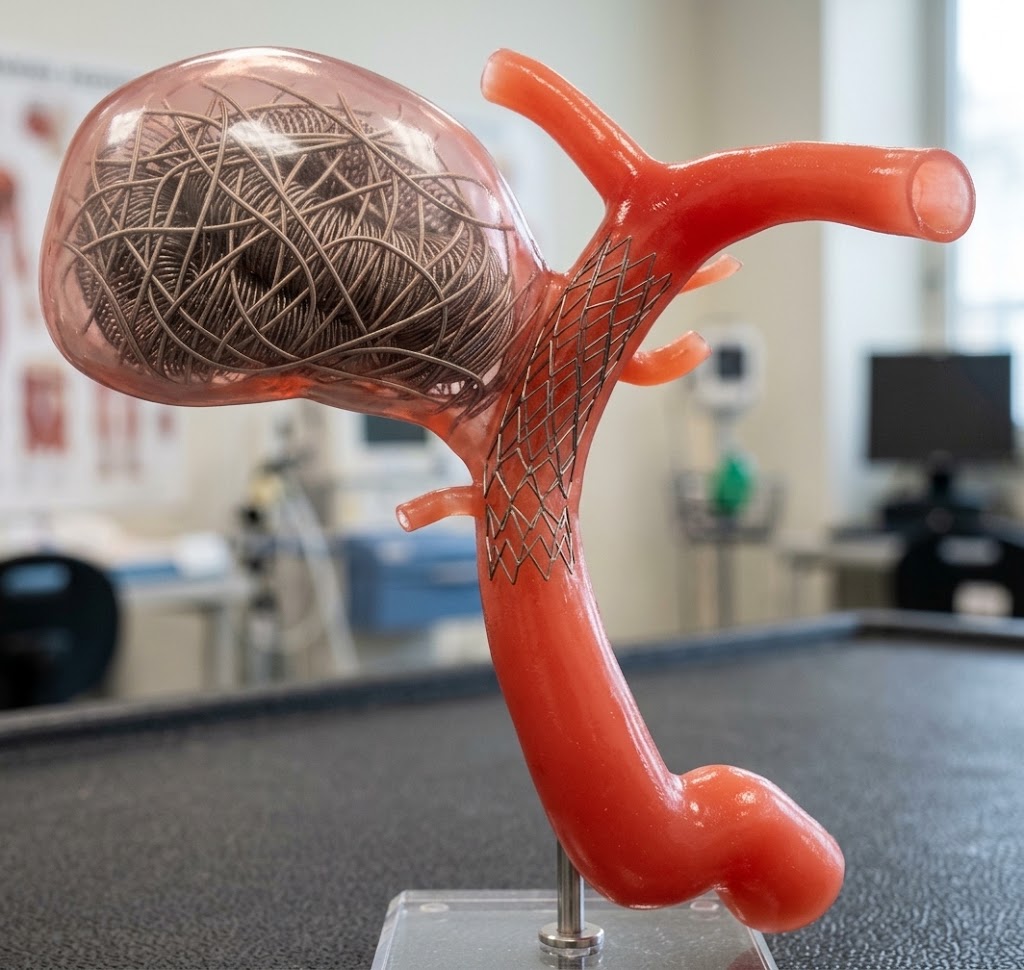
Whether an aneurysm is better treated endovascularly or by microsurgical clipping depends on its location, shape, size, the patient’s age and general health. This decision is best made jointly by a neurosurgeon and an interventional neuroradiologist.
Arteriovenous malformations and fistulas
Embolization delivers liquid embolic agents to close abnormal vessels. In dural arteriovenous fistulas it is often curative on its own. In brain AVMs it is more often part of a combined strategy, reducing blood flow before microsurgery or radiosurgery.
A recent case from our team
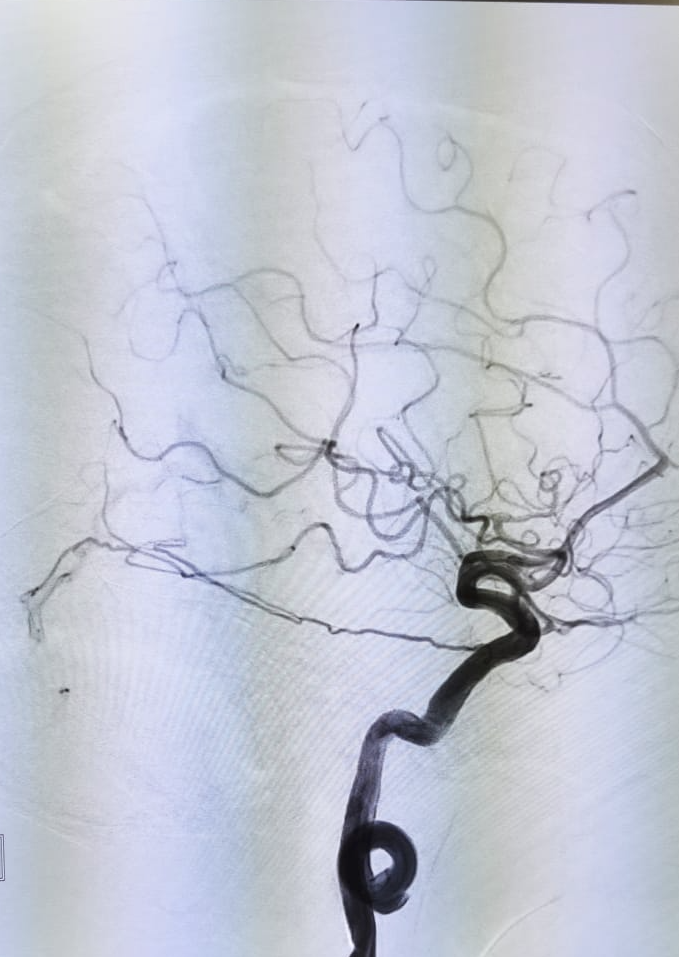
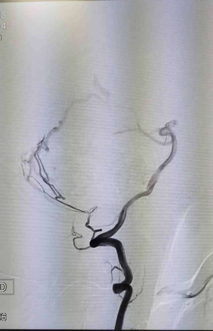
A dural arteriovenous fistula is an abnormal direct connection between an artery and a vein in the dura, the membrane that covers the brain. Fistulas at the tentorium are considered high-risk because they tend to drain into the veins on the surface of the brain, which raises the risk of brain haemorrhage. In this case, the feeding tentorial artery was reached through a microcatheter from inside the vessel and embolized, closing the abnormal connection without open surgery.
Pre-operative tumour embolization
Some tumours, such as certain meningiomas and highly vascular spinal metastases, have a rich blood supply. Closing their feeding vessels a day or two before surgery can reduce blood loss and make tumour resection safer.
Chronic subdural haematoma
Middle meningeal artery embolization is a newer technique for chronic subdural haematoma. Recent randomised trials have shown it reduces the risk of recurrence when added to standard treatment in appropriately selected patients.
A recent case from our team
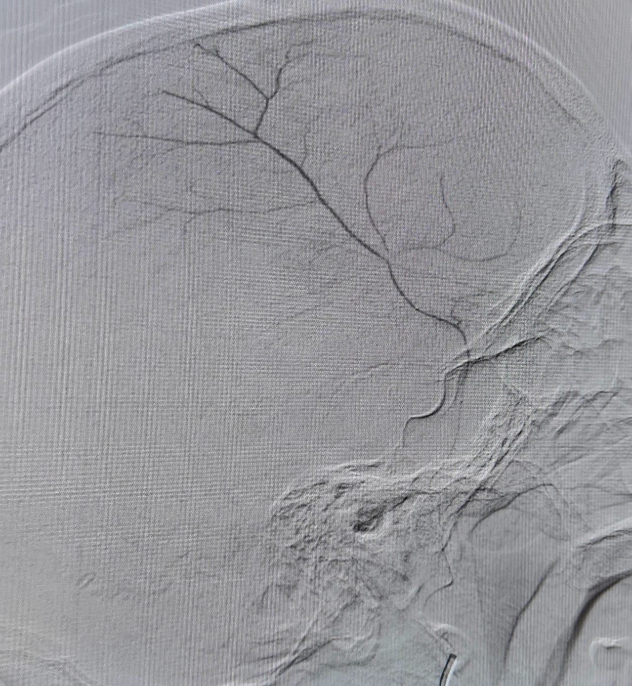
The image shows the middle meningeal artery and its branches, which run along the inner surface of the skull. In chronic subdural haematoma, fragile new vessels arising from these branches keep feeding the collection and make it more likely to come back. Closing them from inside the artery, through a small puncture at the wrist or groin, cuts off that supply and helps the haematoma resolve over the following weeks.
Acute ischaemic stroke
Mechanical thrombectomy removes a clot from a large brain artery and is one of the most effective treatments in modern medicine for major stroke, when performed quickly. Time is critical: anyone with sudden facial droop, arm weakness or speech difficulty should call emergency services immediately (112 in Spain).
A recent case from our team
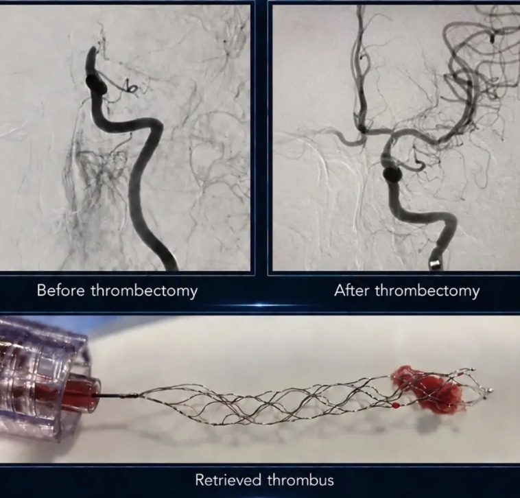
Before treatment, the angiogram shows a major brain artery blocked by a clot, with no blood flow reaching the territory beyond it. A stent retriever was guided through the clot from inside the artery and withdrawn, removing the thrombus in one piece (bottom image). The control angiogram after thrombectomy shows the artery open again and blood flow restored to the brain.
Other indications
Endovascular techniques are also used for carotid and intracranial artery stenosis, cerebral vasospasm after aneurysm rupture, and venous sinus stenting in selected patients with idiopathic intracranial hypertension.
Benefits and limitations
Advantages of the endovascular approach include no skull opening, a smaller wound, usually a shorter hospital stay and a faster return to daily life.
Limitations also exist. Not every lesion can be reached or treated safely from inside the vessel. Some aneurysms are more durably treated with clipping, and some treatments need imaging follow-up or a second session. Patients treated with stents or flow diverters usually need antiplatelet medication for a period afterwards.
Risks are low in experienced hands but not zero. They include bruising or bleeding at the puncture site, reactions to contrast and, rarely, stroke or vessel injury. Your specialist will discuss the specific risk for your case.
Why the combined approach matters
The best results come when endovascular and open techniques are not competitors but tools in the same hands. At Neurospine International, neurosurgery and interventional neuroradiology work together. Each case is reviewed jointly, so the recommendation reflects what is best for the patient, not what a single technique can offer.
For some patients the answer is endovascular treatment. For others it is microsurgery, radiosurgery, a combination, or careful observation. What matters is that the decision is made with the full picture.
When to seek an opinion
You may benefit from a specialist assessment if you have:
- An aneurysm or vascular malformation found on a scan
- A brain haemorrhage of unclear cause
- A recurrent chronic subdural haematoma
- Progressive leg weakness or numbness without a clear spinal explanation
- A tumour planned for surgery that is known to be highly vascular
Neurospine International offers specialist consultations in Marbella and Algeciras, in English and Spanish, for local residents and international patients. Contact us to arrange an appointment.
This article is for general information and does not replace an individual medical consultation.